橫隔膜功能之於核心穩定-上
DIAPHRAGM FUNCTION FOR CORE STABILITY
By Hans Lindgren DC, 9 Jul. 2011
簡介
良好的核心穩定須仰賴橫膈膜執行呼吸以及姿勢維持的雙功能來維持。DNS的創始者Kolar等學者的研究顯示,橫膈膜是重要的姿勢穩定肌肉,而且是可以被自主控制,同時也可以自發性的執行呼吸與姿勢穩定的功能。
然而,近代的核心穩定運動理論中,橫膈膜幾乎根本沒有被提及。在許多專家提供的核心穩定概念中,都完全沒有提到橫膈膜。雖然橫膈膜在近年開始被提到了。可惜的是,仍只有運動處方的最後才強調,在運動過程中要保持橫膈呼吸。
In “core stability from the inside out”we established that proper core stabilization is generated through the diaphragm’s dual function of respiration and postural support. Kolar et al (7) (8) and many others (see references for “Core stability from the inside out”) have shown that the diaphragm is an important muscle for postural stabilization, and also that it is under voluntary control and can perform its respiratory function and postural tasks simultaneously.
It was not that long ago when the diaphragm almost never even got mentioned in the discussions of core and core-stability training. There are still many “experts” who give advice regarding core-stabilization but completely fail to mention the diaphragm. There has however, been more frequent mentioning of diaphragm breathing lately. Unfortunately it often only gets added to exercise prescriptions as final comment of “make sure to maintain diaphragm breathing while exercising”.
何謂橫膈呼吸?
人是無法不依靠橫膈呼吸的。不論你想不想,所有的呼吸都是需要藉由橫膈膜完成,除非你有些隱疾,讓你無法使用橫膈。在一般的呼吸容積中,橫膈的作用約占了80%。
What is diaphragm breathing?
You cannot avoid using your diaphragm when breathing even if you try to! All breathing is performed by the diaphragm whether you want to or not unless there is a medical condition preventing you from using it. The diaphragm is responsible for about 80% of all the respiratory work in normal tidal breathing.
橫膈的解剖和肌動學
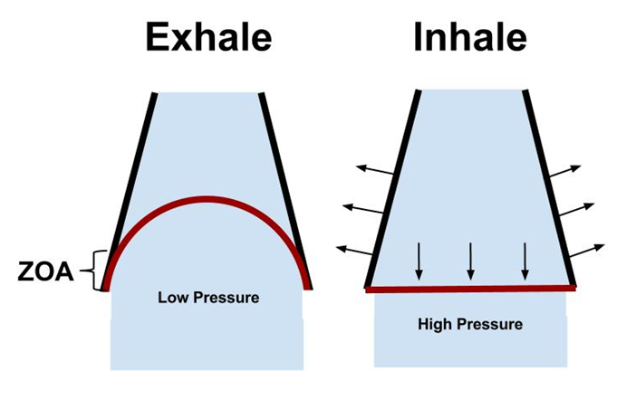
橫膈膜是一個圓頂形狀的肌肉;分隔胸腔與腹腔。中間拱形部分是無法收縮的肌腱,往下會輻射狀的連接到胸廓下段內側。橫膈膜的肋骨端連接到劍突及最後6根肋骨及肋軟骨內側。橫膈膜腳纖維從拱形韌帶延伸連接到上段腰椎。右邊的橫膈膜腳連接到腰椎一到三節,左邊連接到腰椎一二節。胸廓下緣與橫膈膜貼合的區域稱為Zone of apposition (ZOA),這個區域對橫膈膜功能非常重要。這個區域受到腹肌控制且影響橫膈的張力。橫膈的位置及與下段胸廓的解剖關係大大的影響了橫膈膜的效能。
Diaphragm
The diaphragm is a dome shaped muscle separating the thoracic and abdominal cavities. It has a non-contractile central tendon (arcuate) from which muscles radiate caudally and outwards to insert into the inner aspect of the lower ribcage. The costal diaphragm inserts into the xiphoid process and the inner surface of the 6 lower ribs and costal cartilages. The crural fibres span from the arcuate ligament and insert into the bodies and discs of the upper lumbar vertebrae. The right crural diaphragm inserts into L 1-3 vertebrae while the left only inserts into L 1-2. The area of attachment (apposition) between the diaphragm and the ribcage is referred to as the zone of apposition (ZOA) which is of great importance for proper diaphragm function. The zone of apposition is controlled by the abdominal muscles and affects diaphragmatic tension. The diaphragm’s efficiency largely depends upon its position and anatomical relationship with the lower ribcage.
橫膈膜與肋骨貼合區域 (ZOA)
ZOA是個堅硬卻面積變化很大的區域。站姿休息時,這個區域占了內側胸廓30%。橫膈腳在吸氣的時候會從這個區域離開胸廓,使得橫膈收縮往下。ZOA在平靜吸氣時會減少15mm,同時橫膈頂端仍會繼續保持原本的大小及形狀。在肺部最大吸氣量時,這個區域會接近0mm。ZOA肌肉縮短是主要造成橫膈膜在胸廓垂直軸向位移的原因。如果ZOA受限,會造成橫膈在胸廓上的呼吸動作減少。
Zone of apposition (ZOA)
The zone of apposition makes up a substantial but varying area of the ribcage. In standing at rest the human ZOA represents about 30% of the total surface area of the inner ribcage (11). The crural part of the diaphragm peels away from the ribcage at the zone of apposition (10) (9) (12) during diaphragm contraction to allow the diaphragm to descend during inspiration. The zone of apposition decreases by about 15mm during quiet inspiration while the dome of the diaphragm almost remains constant in shape and size. At maximum inspiratory capacity of the lungs the ZOA is almost zero. The shortening of the apposed muscle fibres are mainly responsible for the diaphragm’s axial displacement during inspiration (2) (6). A smaller zone of apposition will result in reduced inspiratory action of the diaphragm on the ribcage (9).
橫膈膜功能
吸氣時,橫膈膜收縮往下,有如活塞進入腹腔內。造成胸腔負壓,讓空氣進入肺,也同時增加了腹內壓。
橫膈是主要的呼吸肌,許多人並沒有正確的活化它。失能的呼吸模式是造成下背痛的常見因素,此因素比其他危險因子要更能預言下背痛的發生。
Diaphragm function
During inspiration the diaphragm contracts and moves down caudally like a piston into the abdominal cavity, which creates a negative pressure in the thoracic cavity that forces air into the lungs and simultaneously increases the intra-abdominal pressure.
The diaphragm is our primary breathing muscle and yet many individuals have very little awareness of how to activate it properly. Dysfunctional breathing patterns are a common contributing factor for low-back pain conditions, and it is actually often a stronger predictor for low back pain than other established risk factors (15).
正確的橫膈膜呼吸與腹式呼吸(belly breathing)一樣嗎?
橫膈膜呼吸一直被視為腹式呼吸,其實未必。當橫膈膜收縮下降到腹腔,腹內壓增加會造成腹部膨脹。有效的橫隔呼吸中,腹壁肌肉的膨脹應該是3D的,所有的方向都輕微的擴張。腹壁所有肌肉會以離心收縮的方式來對抗橫膈所給予的壓力。腹壁肌肉對抗橫膈膜的能力同時也對橫膈膜長度與張力之間是非常重要的控制。
骨胳肌中,包括橫膈都有長度與張力之間的關係,即長度縮短後(收縮),收縮的力量也隨之下降。這股由腹壁肌肉離心收縮製造的力量可以維持橫膈膜與肋骨貼合的區域以及橫膈圓頂的形狀。進而促進了橫膈力量的增加。腹式呼吸只有擴張腹腔的前半部並沒有提供橫膈膜任何的阻力,事實上,還減少了橫膈有效收縮的能力。
Is proper diaphragm breathing the same as belly breathing?
Diaphragm breathing is often referred to as belly breathing, but that is not correct. When the diaphragm contracts and descends into the abdominal cavity the intra-abdominal pressure increases and will distend the abdominal wall. In efficient diaphragm breathing the distension of the abdominal wall should be three dimensional with a slight expansion in all directions. The abdominal wall should oppose the action of the diaphragm with an eccentric contraction of all the abdominal muscles. The opposing action of the abdominal wall is very important in controlling the length tension relationship of the diaphragm muscle.
Any skeletal muscle, including the diaphragm, has a length-tension relationship where decreased length (contraction) decreases the force of the contraction. The opposing forces created by the abdominal muscles in their eccentric contraction maintain the zone of apposition and the dome shape of the diaphragm, and thereby facilitates the increased force of the diaphragm. Belly breathing only distends the abdomen forward, which does not offer any resistance to the diaphragms motion and will therefore actually reduce the diaphragm’s ability to contract efficiently.
理想的橫膈膜呼吸
理想的橫膈膜呼吸主要是下肋骨側方的擴張。橫膈膜收縮時,肋骨端橫膈(ZOA)的收縮會擴張下段胸廓以及腹壁,橫膈膜腳端,由於解剖走向連接到腰椎,收縮時,只會造成腹部往前的方向。ZOA可以將腹內壓傳送到胸廓。形成吸氣時,橫膈膜收縮影響胸廓使其往外的機制。這是橫膈膜在連接胸廓處對肋骨形成往外、往上的動作(外旋)。
Proper diaphragm breathing
Ideal diaphragm breathing expands the lower ribs outwards in a mainly lateral direction. The costal part of the diaphragm (ZOA) expands both the lower ribcage and the abdominal wall when contracting (the crural part only displaces the abdomen forward with its contraction directed forward due to its attachments on the lumbar spine). The apposition of the diaphragm to the inner ribcage wall allows for transmission of intra-abdominal pressure to the ribcage, which provides a mechanism whereby the diaphragmatic contraction drives the ribcage outwards during inspiration (16) (11). Adding to this is the direct outward lifting motion (external rotation) the diaphragm exerts on the ribs at its insertions to the ribcage.
良好橫隔膜呼吸的特徵
下段胸廓應該要有擴張而且胸腔不會往頭側移動。伴隨著腹部肌肉協同做輕微擴張動作,並透過離心收縮來控制腹內壓。
Signs of proper diaphragm breathing
There should be an expansion of the lower ribcage without any cranial movement of the chest, accompanied by a synchronized activity of the entire abdominal wall which expands slightly while controlling the IAP by an eccentric contraction.
沒有留言:
張貼留言